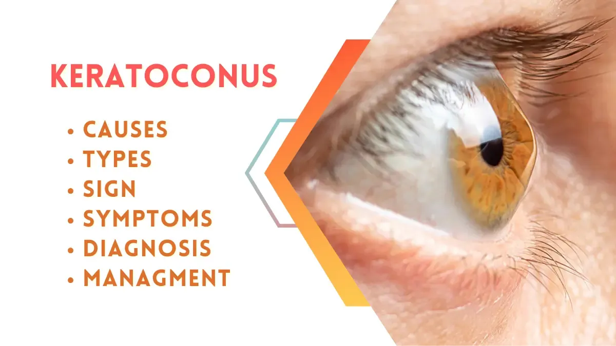
Keratoconus
Keratoconus is a progressive eye disorder where the normally round cornea (the clear front part of the eye) thins and bulges into a cone-like shape. This cone shape deflects light as it enters the eye, causing distorted vision. Keratoconus affects roughly one in every 2,000 people, usually starting during puberty and continuing to progress until the mid-30s.
Definition
Keratoconus is a non-inflammatory, degenerative condition of the cornea characterized by localized thinning, which leads to irregular astigmatism and vision distortion.
Types of Keratoconus
- Forme Fruste Keratoconus: A very mild, non-progressive form often detected during corneal topography.
- Mild Keratoconus: Slight thinning and distortion of the cornea with minor vision impairment.
- Moderate Keratoconus: Noticeable vision distortion that often requires corrective lenses.
- Severe Keratoconus: Significant thinning with marked cone-shaped protrusion, often requiring surgical intervention.
- Acute Keratoconus (Hydrops): Sudden swelling of the cornea due to a tear in Descemet's membrane, causing vision loss and pain.
Causes
- Genetic Factors: Family history of keratoconus increases risk.
- Chronic Eye Rubbing: Common in people with allergies or dry eyes.
- Underlying Conditions: Associated with Down syndrome, Ehlers-Danlos syndrome, and Marfan syndrome.
- Oxidative Stress: Imbalance of free radicals and antioxidants in the cornea.
- Environmental Factors: Prolonged exposure to UV light.
Signs and Symptoms
- Blurred or distorted vision.
- Increased sensitivity to light (photophobia).
- Frequent changes in eyeglass prescription.
- Double vision in one eye (monocular diplopia).
- Halos and glare around lights.
- Eye strain or discomfort.
- Vision difficulty in low-light conditions.
Diagnosis
- Visual Acuity Test: Checks clarity and sharpness of vision.
- Slit Lamp Examination: Examines the cornea for thinning or scarring.
- Corneal Topography: Maps the shape and curvature of the cornea to detect irregularities.
- Pachymetry: Measures corneal thickness.
- Ophthalmoscopy: Evaluates the overall health of the eye.
Investigations
- Pentacam Imaging: Detailed 3D imaging of the cornea.
- Keratometry: Measures corneal curvature. Normal corneal curvature (K) values range between 40-48 diopters (D). In keratoconus, the values may exceed 48 D, often reaching 50-60 D or more in severe cases.
Treatment
1. Non-Surgical Treatments
- Glasses or Soft Contact Lenses: For mild cases. If someone has Keratoconus, usually the power of the eyes goes towards minus (myopia). Along with this, astigmatism also increases, which is not regular (irregular astigmatism).
- Rigid Gas-Permeable (RGP) Lenses: Provide better vision correction in moderate cases.
- Scleral Lenses: Cover the entire cornea for severe cases.
- Corneal Collagen Cross-Linking (CXL): Stabilizes the cornea by strengthening collagen fibers.
- Medicine used in CXL: Riboflavin (Vitamin B2) combined with UV light.
2. Surgical Treatments
- Intacs: Implantation of small corneal rings to flatten the cone.
- Corneal Transplant (Keratoplasty): For advanced cases with severe thinning or scarring.
- Topography-Guided PRK: Reshapes the cornea using a laser.
Medicines for Symptom Management
- Lubricating Eye Drops: Carboxymethylcellulose or hydroxypropyl methylcellulose drops for dry eyes.
- Anti-Allergy Drops: Olopatadine or Ketotifen for eye allergies.
- Steroid Eye Drops: Fluorometholone or Loteprednol for inflammation (short-term use).
Medicines to Avoid
- Corticosteroids (Long-Term Use): Prolonged use may exacerbate corneal thinning.
- NSAID Eye Drops: Can delay corneal healing.
Role of Vitamins
- Vitamin A: Improves eye surface health. Found in carrots, sweet potatoes, and spinach.
- Vitamin C: Supports collagen production, vital for corneal strength.
- Vitamin E: Reduces oxidative stress.
- Omega-3 Fatty Acids: Decrease inflammation and improve tear quality.
Lifestyle Effects
- Difficulty driving, especially at night.
- Challenges with reading or working on computers for extended periods.
- Increased dependence on visual aids.
- Psychological impact due to vision impairment.
Complications
- Corneal Scarring: Causes further vision loss.
- Hydrops: Sudden swelling leading to severe pain and vision loss.
- Keratoplasty Failure: Rejection of a corneal transplant.
Management
- Regular Monitoring: Routine eye check-ups for progression.
- Protective Eyewear: Prevents UV damage.
- Avoid Eye Rubbing: Reduces risk of progression.
- Hydration and Diet: Promote eye health with a balanced diet.
- Psychological Support: Address anxiety or depression due to vision challenges.
By understanding keratoconus in-depth, patients and caregivers can manage the condition effectively and improve quality of life. Always consult an ophthalmologist for personalized care and treatment.

