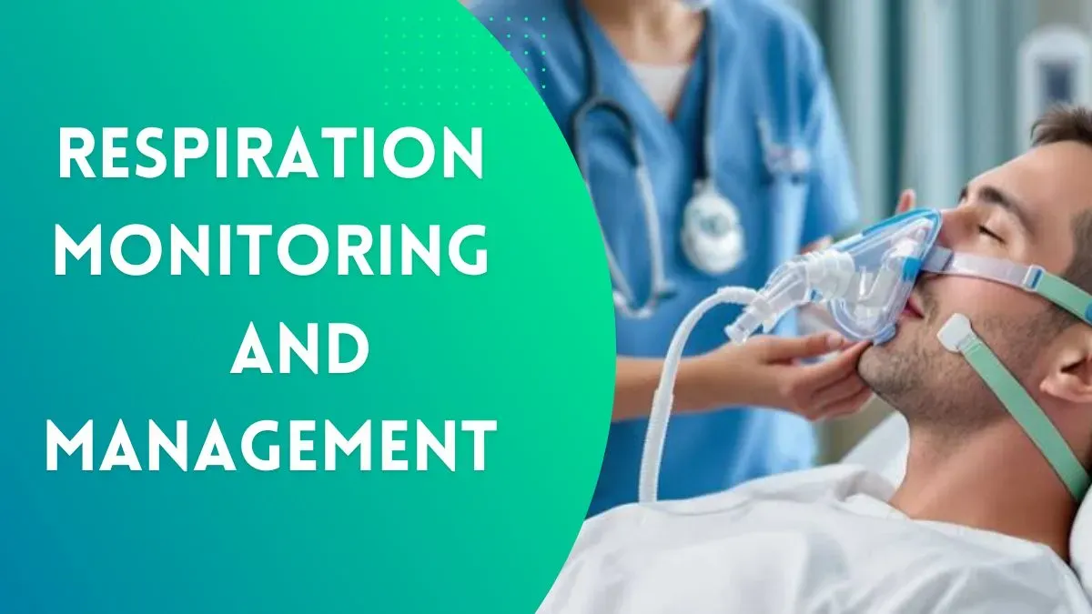
Respiration Monitoring
Respiration monitoring is an essential aspect of clinical practice, allowing healthcare professionals to assess a patient's breathing patterns, oxygenation, and overall respiratory function.
It is particularly important in critical care, anesthesia, emergency medicine, and pulmonary medicine.
Physiology of Respiration
Respiration involves two key processes:
- External Respiration - Exchange of gases (O2 and CO2) between the lungs and the blood.
- Internal Respiration - Exchange of gases between the blood and tissues.
The respiratory rate is controlled by the medullary respiratory center, which responds to changes in carbon dioxide (CO2) levels, oxygen (O2) levels, and pH.
Respiration Monitoring Parameters
Respiratory monitoring includes the following key parameters:
- Respiratory Rate (RR): Normal range is 12–20 breaths per minute in adults.
- Tidal Volume (TV): Volume of air moved per breath (~500 mL in adults).
- Minute Ventilation: Product of RR and TV.
- Oxygen Saturation (SpO2): Measured using pulse oximetry (normal > 95%). Normal SpO2 range is 95% - 100%.
- End-Tidal CO2 (ETCO2): Measures exhaled CO2 to assess ventilation.
Respiration Monitoring Methods
1. Clinical Examination
- Inspection: Observing chest movements, use of accessory muscles.
- Palpation: Checking for symmetry of chest expansion.
- Percussion: Identifying areas of hyperresonance or dullness.
- Auscultation: Listening for breath sounds, wheezing, crackles, or stridor.
2. Pulse Oximetry
- Non-invasive method measuring oxygen saturation (SpO2).
- Uses infrared light to assess hemoglobin oxygenation.
- Limitations: Affected by poor perfusion, nail polish, and motion artifacts.
3. Capnography
- Measures end-tidal CO2 (ETCO2) using a nasal or endotracheal sensor.
- Provides real-time feedback on ventilation and metabolic status.
- Normal ETCO2: 35-45 mmHg.
4. Arterial Blood Gas (ABG) Analysis
- Provides detailed information on pH, PaO2, PaCO2, and bicarbonate levels.
- Helps assess respiratory acidosis/alkalosis and oxygenation status.
5. Imaging Techniques
- Chest X-ray (CXR): Assesses lung pathology (e.g., pneumonia, pleural effusion, pneumothorax).
- Computed Tomography (CT): Provides detailed images of lung structure.
- Ultrasound: Used for diagnosing pleural effusion and lung consolidation.
6. Electronic Respiratory Monitors
- Devices such as impedance pneumography and wearable sensors track respiratory patterns.
- Used in ICU and home care settings.
Clinical Applications
- Critical Care: Continuous monitoring in ICU patients with respiratory failure.
- Anesthesia: Ensuring adequate ventilation during surgery.
- Emergency Medicine: Rapid assessment in trauma, asthma, or COPD exacerbations.
- Pulmonary Diseases: Monitoring in conditions like pneumonia, ARDS, and sleep apnea.
Interpretation & Abnormal Patterns
Tachypnea: RR > 20 breaths/min (e.g., fever, sepsis, metabolic acidosis).
Bradypnea: RR < 12 breaths/min (e.g., opioid overdose, brainstem injury).
Cheyne-Stokes Breathing: Alternating periods of apnea and hyperventilation (e.g., heart failure, stroke).
Kussmaul Breathing: Deep, labored breathing (e.g., diabetic ketoacidosis).
Apnea: Absence of breathing (e.g., sleep apnea, respiratory arrest).
Conclusion
Respiration monitoring is a crucial aspect of patient assessment and management. It provides vital information about a patient’s respiratory and metabolic status, guiding clinical decisions in both emergency and chronic care settings. Understanding the principles and techniques of respiratory monitoring is essential for medical students and healthcare professionals preparing for exams and clinical practice.

